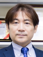
Prof. Kenji Suzuki
Institute of Science Tokyo, Japan
Kenji Suzuki, Ph.D. worked at Hitachi Medical Corp, Aichi Prefectural University, Japan, as a faculty member, in Department of Radiology, University of Chicago, as Assistant Professor, and Medical Imaging Research Center, Illinois Institute of Technology, as Associate Professor (Tenured). He is currently a Full Professor (Tenured) & Founding Director of Biomedical Artificial Intelligence Research Unit, Institute of Integrated Research, Institute of Science Tokyo, Japan. He published more than 395 papers (including 125 peer-reviewed journal papers). He has been actively researching on deep learning in medical imaging and AI-aided diagnosis in the past 25 years, especially his early deep-learning model was proposed in 1994. His papers were cited more than 16,000 times, and his h-index is 63. He is inventor on 37 patents (including ones of earliest deep-learning patents), which were licensed to several companies and commercialized via FDA approvals. He published 16 books and edited 20 journal special issues. He has been awarded numerous grants including NIH, NEDO, and JST grants, totaling $8M. He serves as Editors of more than 20 leading international journals including Pattern Recognition and AI. He chaired 110 international conferences. He is a Fellow of IARIA. He received 25 awards, including 3 Best Paper Awards in leading journals.
Speech Title: "Small-Data AI for Diagnosis of Rare Lesions in Medical Images and Medical AI Imaging"
Abstract: Artificial intelligence (AI) basdd on deep learning has shown to be a breakthrough technology in many fields including medicine. My group has been actively studying on deep learning in medical imaging in the past 25 years, including ones of the earliest deep-learning models for medical image processing, lesion/organ segmentation, and classification of lesions in medical imaging. In this talk, "small-data" AI that can be trained with a small number of cases/images is introduced. A chief limitation of current deep learning models is the requirement of a huge number of images/cases, "big data", to train. To address this issue, we succeeded in developing small-data deep learning model that does not require "big data". Our small-data AI was applied to develop AI-aided diagnostic systems (“AI doctor”) and deep-learning-based imaging for diagnosis (“virtual AI imaging”), including 1) AI systems for detection and diagnosis of lung, colon, breast, liver, and rare cancer in medical images, and 2) virtual AI imaging systems for separation of bones from soft tissue in chest radiographs and those for radiation dose reduction in CT, tomosynthesis, and mammography. Some of them have been commercialized via FDA approval in the U.S., including the first FDA-approved deep-learning product. Our small-data deep-learning technology would be useful for the development of AI in “small-data” areas where “big data” are not available.

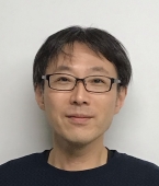
Prof. Yoshiaki Yasuno
University of Tsukuba, Japan
Yoshiaki Yasuno leads
Computational Optics Group at the University of Tsukuba. He obtained
his PhD based on his research work of spatio-temporal optical
computing in 2000 and extend this conception to optical measurement
including Fourier-domain optical coherence tomography (OCT). Since
2003 he has been working for ophthalmic OCT imaging, including
retina and anterior eye. He has also actively worked for OCT
angiography and polarization sensitive OCT. His current research
interests cover multi-functional optical coherence microscopy, its
augmentation by using computational technologies, and theoretical
modeling of modern metrology.
Speech Title: "Label-free Metabolic Imaging of in Vivo Samples by Computationally Augmented Optical Coherence Microscopy""
Abstract: Recent advancements in cultivation technology enable thick and functional in vitro samples, which enable effective assessment of drugs and chemical compounds. Despite they become increasingly important, suitable imaging technology for these samples is still missing. Optical coherence tomography (OCT) based microscopy (OCM) has been used for these samples and has enabled non-invasive and volumetric imaging. However, OCM cannot assess the dynamic functions of the tissue. Here we present several computational extensions of OCM, which overcome these problems. Namely, time- statistics analysis enables intracellular-dynamics-based contrast. And a neural-network-based extension accelerates the overall measurement speed of OCM.

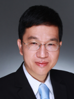
Prof. Rongshan Yu(SMIEEE, FIET)
Vice Director, National Institute for Data Science in Health and Medicine, Xiamen University
Dr. Rongshan Yu received his bachelor degree in Electrical Engineering with minor in Applied Mathematics from Shanghai Jiaotong University, China in 1995, and the PhD degree from the National University of Singapore (NUS, Singapore) in 2004. He is currently with the Department of Computer Science, Xiamen University as a professor, and vice-director of the National Institute for Data Science in Health and Medicine, Xiamen University. His research interests include high-throughput multi-omics data analysis, precision medicine, and medical artificial intelligence. He has more than 180 journal/conference publications and holds more than 20 US/International patents. Dr. Yu was a member of the Technical Committee of the Multimedia Systems and Applications of the IEEE Circuits & Systems Society, and chair of Internet of Thing (IoT) Special Interest Group (SIG) of IEEE Signal Processing Society. He served as standard project editors of ISO/IEC 14496-3/AMD.5 (MPEG Audio Scalable Lossless Coding) and ISO/IEC 14496-5:2001/Amd.10 (SSC, DST, ALS, and SLS reference software), and associated editors of IEEE Transactions on Audio, Speech and Language Processing and IEEE Access. He is a senior member of the Institute of Electrical and Electronics Engineers (IEEE), and Fellow of the Institution of Engineering and Technology (IET). In 2007, he received the National Technology Award, Singapore in 2007 in recognition of his contributions to international standards.
